


Arthrosis is a whole group of distrust depth of joint equipment, with various etiology, but a similar clinical picture of abnormal changes.The jointin cartilage of the joint, then the drunken bone tissue, the joint capsule and the league, are exposed to destruction and deformation.The disease is chronic progressive and can significantly limit the patient's motor activity without proper treatment.
Diagnosis and treatment of pathology is an arthrologist, rheumatologist, surgeon, orthopedic.
General information
Arthrosis is diagnosed with about 1/5 of the planet's population, but the disease is more typical of elderly.This proves the statistics of spread between different ages:
- Young people last for up to 40 years to more than 6-7 %;
- Mature faces after 45 years to 20-25%;
- After 70 years - up to 80%.
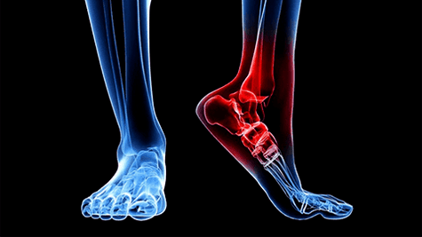
The disease affects tissues under constant load: small joints and feet, hips and knee joints, cervical and chest parts of the spine, a little less frequently ankle and shoulder joints.
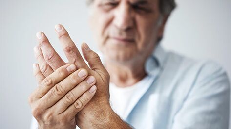
Note! The defeat of the interfacing joints of the hands is ten times more common in women than in men.
In many Western countries, the term "arthrosis" is not used, replacing it with the concept of "arthritis".Such a substitution is quite justified as inflammatory processes in most cases prevent arthrosis or escort.In home medicine, the terms "arthrosis" and "arthritis" are essentially the same disease, but with different etiology of the process.In addition, osteoarthritis, osteoarthrosis, deforming osteoarthrosis are used to denote pathology.
Note! The difference between arthritis and arthrosis lies in the cause of the disease.In the first case, these are inflammatory processes (meaning of suffix inflammation) in the second -metabolic disorders (protein, mineral).
The mechanism of development and joint reasons
The main cause of arthrosis is a violation of anabolism and cartilage and bone tissue catabolism.If the synthesis processes are normally prevalent, the death processes will go faster during the changes in the Arthrose.As a result, we observe the rapid aging and degeneration of tissue structures.First they start to collapse at the cellular level and then on the organ tone.The first devastating changes appear:
- the accumulation of cartilage;
- superficial storytelling;
- micro -clots and tears;
- Focus and general thinning of the cartilage layer.
The cartilage loses natural elasticity and density and is no longer able to serve as a shock absorber during movements.The mutual correspondence of the shape of the joint surfaces is disturbed, leading to deformation of the relationship.This exacerbates the development of abnormal changes and causes many irreversible processes.In exchange for lost cartilage, bone tissue begins to grow with spikes and outgrowths, which can lead to severe disability of fetter movements and later the patient.
The reasons for this scenario are:
- Violation of mineral metabolism can lead to gout changes in the joints, osteoporosis, etc.
- The disadvantage of tissue nutrition is venous stagnation and poor microcirculation slows down blood supply and lymphatic drainage.The mineral composition of the bone is exhausted, becomes osteoporous and loses suicide ability.The phenomenon is characterized by vascular pathologies and hormonal failures.
- Inflammatory processes - acute infectious diseases, body hypothermia, impaired hormonal background.
- Autoimmune reactions Chronic focuses of inflammation, nervous stress, endocrine pathologies and other causes can provoke the body's immune system aggression against its own cells, including joint tissues.The most common autoimmune lesions rheumatoid arthritis, scleroderma and red lupus.
- Increased joint wear - the deviation between functionality and deposited load slows down the synthesis processes and accelerates destruction.The phenomenon is characterized by athletes, dancers, overweight people, and anyone who is dealing with heavy physical work or is associated with long static (constant work).
- Injuries - bruises, dislocations, fractures, penetrating wounds, tears - violate the structure of the tissues and give impetus to the beginning of deformation.
- Genetically defined pathologies - connective tissue dyslasia, violation of collagen synthesis initially form an unstable, low functional joint.
Some reasons echo tightly with each other and form a complex abnormal complex.
Attention! Hormonal differences play a particularly important role in violation of bone tissue metabolism.Taking thyroid failure, menopause, contraceptives, corticosteroids - this is a direct way for osteoporous and arthrose changes in the skeleton.
Classification of changes
There are many major criteria for arthrosis systematics: causes and etiology, localization, coverage.
Etiology:
- Primary arthrosis - develops independently with the damage to completely healthy joints without the participation of previous pathologists;
- It is in secondary, in the background of the disease (gout, psoriasis, rheumatism) and in the presence of existing joint deformations or injuries.
According to the degree of coverage:
- Local shapes for a limited number of joint damage: Monoarthrosis-1 joint, oligoarthritis-2-3;
- The general shapes of various types of polyarthrosis when 3 large structures and many others are involved in the pathological process.
According to the process, the name of the arthrosis of each joint is given separately:
- Coksartrosis - disables hip connection;
- Spondylarthrosis - intervertebral discs, primarily the cervix, chest and groin;
- Gonarthrosis - with harmful work of the knee joint;one of the most common species;
- Cruzartrosis - participating in the ankle process.
Arthrosis can be progressive, compensated or decompensated quickly or slowly.
The main symptoms and signs
Arthrosis is a complex disease.Traditionally, it can be divided into several pathologies:
- Chondritis and chondrosis - inflammatory and degenerative lesions of cartilage tissue;
- Osteoporosis is also osteoporosis - a pathological process in bone structures;
- Synovitis - involving the lining of the joint capsule;
- Bursit - overall inflammation of the joint bag;
- Reactive damage to the soft tissues of the adjacent area - affects muscles, ligaments, fiber.
They can be observed simultaneously or selectively depending on the stage, degree and shape.With this in mind, the complex of symptomatic changes develops.Including:
- Pokhrutzhazing is a symptom of damage to mineral metabolism and the first sign of the disease.Can occur at any age.
- Set - intensively opening in the morning.It is short -term and can be expressed as a result of the joint jam.
- Restriction of mobility - reducing the amplitude of movements in the Committee of Active or Passive Acts.
- Pain-relieving manifestation, starting from unpleasant and sore, which after intense loads obtains a background figure and acute sharp when it ends with executive movements.The so -called "starting pains" are particularly typical, which are manifested after a long period of rest and last until the joint is completely formed.
- Swelling - inflammation of the soft tissues, synovitis, bursitis.
- Deformation - is observed with the complete degeneration of the cartilage and the lack of shock abstitlation.
Note! Bushara and Geberden knots are a typical sign of deforming arthrosis of the hand.These are bone growth with the processes of osteophytes.
The joint sections and degrees
The intensity of joint changes can be distinguished by the 4 stages of the disease:
- Section 1 - with a slight change of cartilage (violation of structure and functionality in the collagen fibers).The image in X -Gray is virtually unseen.
- Stage 2 - Capial tissue pinch can reach up to 50%in the lumen of the joint.It is covered with cracks, with slight pain in the damaged compound.Osteophytic complexes appear on X -try;The joint scatter slightly reduces its size.
- Stage 3 - The lesion of the cartilage almost reaches the base of the bone, and the joint gap sharply decreases.
- 4 Stage - The cartilage is completely damaged, leading to partial or complete degeneration of the synovial fluid, bone tissues with each other and deformation of the compound.In some areas, changes in sclerosis develop.Extreme manifestation of arthrosis is the merger of joint tissues with the bone of the structures and the total loss of mobility.
In some sources, stages 1 and 2 combine.
As symptoms progress, a person's motor activity suffers.Given the functional performance of the joint, 4 degrees of possible development of pathology are distinguished:
- 0 degrees - mobility and performance remain in full;
- 1 degree - the patient retains the ability to manifest itself and social activity, but is unable to participate in the workforce;
- 2 degrees - the difficulties of the manifestation of social activity add to the violation of the workforce;
- 3 degrees - all types of activities are limited or completely impossible: work, social and self -service;The patient needs constant care.
What are the potential complications
By tightening treatment you can provoke many unpleasant consequences:
- constant pain syndrome;
- lameness;
- vertebrates (with spondylarthrosis);
- pronounced joint deformation;
- Full mobility with the ossification of structures.
What does the survey procedure contain
A medical examination is sufficient to diagnose arthrosis by collecting a history.An instrumental examination is performed to determine the degree of injury.The main way to obtain a clear image of the disease is:
- radiography;
- CT, MRI;
- ultrasound;
- scintigraphy;
- Diagnostic arthroscopy with biopsy of cartilage tissue and synovial fluid.
In an acute inflammatory procedure, the doctor prescribes additional analysis: general blood test, rheumens, biochemistry (glucose, protein compounds, electrolytes).
Treatment
It is impossible to complete the disease.Timely treatment of arthrosis allows you to maintain the functionality of joint, normal motor activity and prevent pain.It should begin in the first stage to exclude complications.
Medication Includes:
- Anti -inflammatory drugs, primarily NSAIDs;
- intraarticular steroid pain and inflammation (pronounced synovitis, bursitis);
- Proteolysis inhibitors - slow down and suspend the process of destruction of bones and cartilage;
- Malispazmodics - prevent muscle cramps;
- angioprotectors and drugs in the affected tissues to improve blood microcirculation;
- chondroprotectors;
- synthetic substitutes for synovial fluid;
- Vitamin and mineral compositions.
The complex of physiotherapy It is prescribed in parallel to improve the effects of the drugs.The main physiotherapy:
- magnetic therapy;
- electrophoresis;
- UHF;
- mud;
- bath;
- massage;
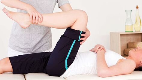
- Practical therapy and kinesiotherapy using special simulators.
Surgery - The only way to treat later stages is when the cartilage tissue is completely destroyed.The following solutions are possible for the problem:
- Endoscopy - partial or complete replacement of the joint with artificial analog;
- Arthroscopy - a minimally invasive operation for removing osteophytes or partial replacement of the cartilage;
- Artrodesis - closure of the joint and fixing it in the most convenient position;The motionless joint surfaces increase over time.
Forecast
Arthrosis does not threaten the patient's life, but lack of treatment can significantly limit freedom of movement and exacerbate quality of life.Timely and competent therapy can restore the healthy condition of the joint at an early stage.In other cases, only a slowdown in the degeneration process is possible to compensate for the functions that are lost due to conservative treatment and prosthetics.
Preventing the disease
Complete healing is almost impossible, so prevention should be paid special attention.The main requirement is to treat healthy lifestyle and inflammatory processes:
- Do not allow hypothermia or treat infectious diseases in time;
- Avoid physical overload and long static loads;
- Maintains normal body weight;
- Contact the proper diet-the balanced composition of vitamins and minerals is very important for the health of the muscle bone;
- Completely (if possible, until full healing), treat any damage to the joints;
- Practice systematic physical exercises to stimulate blood circulation (bicycle, hiking, light jogging, Scandinavian walk).
If you are in danger (elderly, poor inheritance, physical overload), be sure to perform regular radiographic examination.